
The feeling is unmistakable. It is that sudden, sickening pop during a pivot, or the creeping, dull ache that starts during a long run and doesn’t stop once you finally sit down. Your knee is the largest joint in your body, and it acts as a complex hinge that handles the entire weight of your body while facilitating everything from simple walking to explosive athletic maneuvers. When that hinge stops working, the rest of your movement patterns fall apart.
Healing a knee injury requires more than just stopping the activity that caused the pain. It demands a methodical approach, balancing the need for immediate tissue protection with the necessity of restoring strength, stability, and range of motion. Ignoring small niggles usually leads to larger, systemic issues because your body will instinctively compensate, shifting stress to your hips, ankles, or lower back.
This guide breaks down fifteen of the most common ways knees fail, why they happen, and exactly how to navigate the road to recovery. You do not need to be a medical professional to understand the mechanics at play, but you do need to be disciplined. Recovery is rarely about finding a magic bullet; it is about patient, consistent loading and restoring the architecture of the joint.
1. Patellar Tendonitis (Jumper’s Knee)
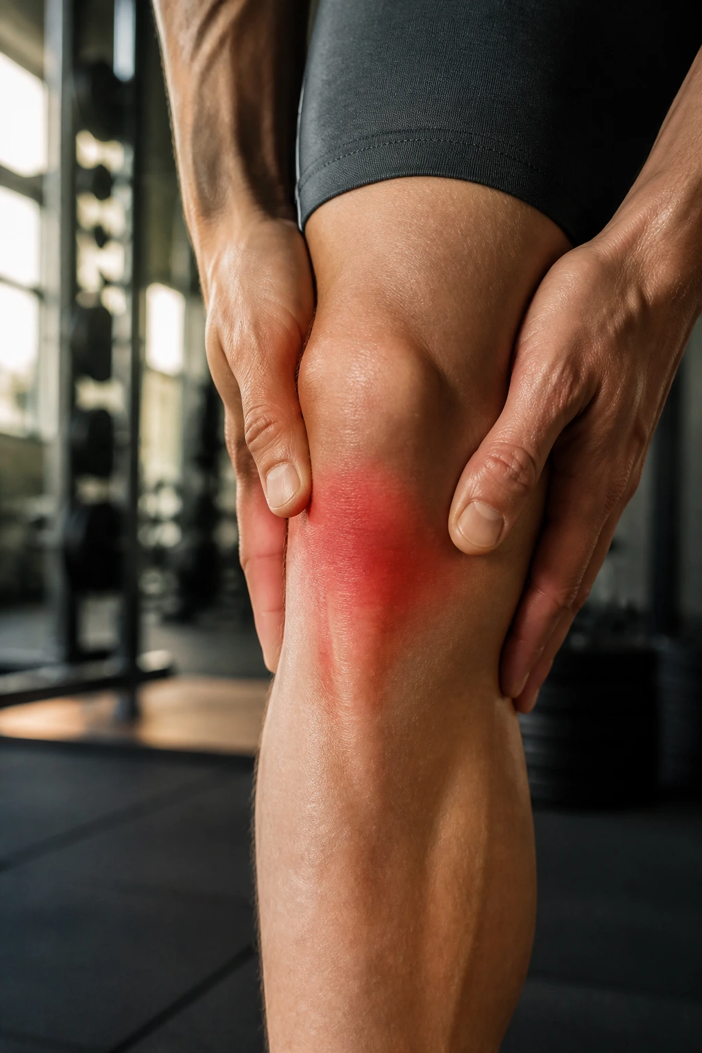
Jumper’s knee is arguably the most annoying injury for athletes who love explosive movement. It is essentially an overuse injury where the tendon connecting your kneecap to your shinbone becomes inflamed, irritated, or—in chronic cases—begins to degenerate. You will feel this right below the kneecap. It often presents as a localized, sharp pain that worsens when you jump, squat, or sprint.
The Mechanics of the Pain
The patellar tendon acts as a spring, absorbing force when you land and releasing it when you jump. If your quadriceps are too tight or your landing mechanics are inefficient, that tendon takes on more load than it can handle. Over time, micro-tears develop faster than your body can repair them, leading to the thickening and pain associated with tendonitis.
How to Heal It
- Load Management: Stop high-impact jumping immediately. You need to offload the tendon to let the inflammation subside.
- Isometric Holds: Research shows that isometric exercise—holding a squat at a specific angle—can actually provide immediate pain relief for tendon issues. Hold a wall sit or a shallow squat for 45 seconds, repeating five times.
- Eccentric Focus: Once the pain calms down, start slow, controlled eccentric movements. Use a decline squat board, lowering yourself over three seconds and coming up with two legs.
- Strengthen the Glutes: Often, the quads are overworked because the glutes aren’t doing their job. Weak glutes mean the knee has to work twice as hard.
Pro tip: Do not stretch your quadriceps aggressively while the tendon is actively flared up. You are pulling on a compromised structure that needs stability, not tension.
2. Anterior Cruciate Ligament (ACL) Tear
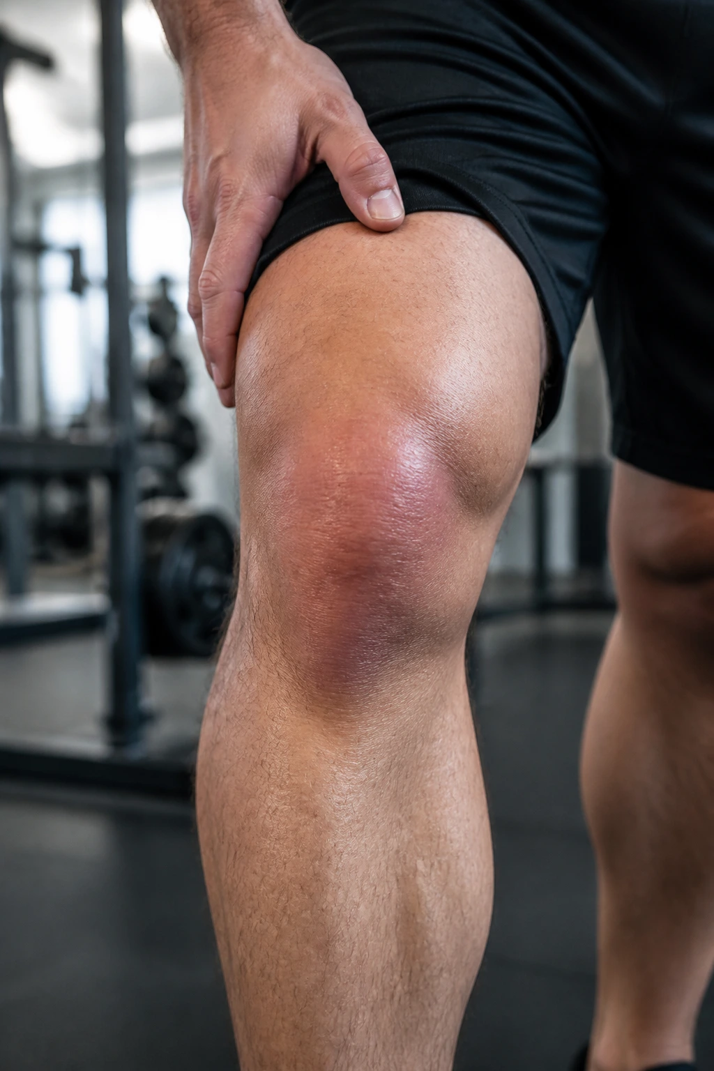
The ACL is the primary stabilizer that prevents your shinbone from sliding out in front of your thighbone. When it tears, it is usually because of a non-contact deceleration or a sudden change of direction where the knee buckles inward. Most people hear or feel a loud pop, followed by rapid, significant swelling and a feeling that the knee is “giving way” whenever they try to put weight on it.
Understanding the Instability
An ACL tear is not just an injury to a single ligament; it changes the entire mechanical integrity of the knee. Without that internal strap, your femur and tibia move against each other improperly, which causes a grinding sensation that can damage the meniscus over time if left unaddressed.
The Healing Path
For many athletes, surgical reconstruction is the standard, but the real work happens in the months before and after the surgery. Pre-habilitation—strengthening the leg before the procedure—significantly improves post-operative outcomes.
Recovery is a marathon, not a sprint. You are looking at a recovery timeline that spans months, focusing on:
- Regaining Full Extension: This is the most critical first step. If you cannot lock your knee out straight, your gait will be compromised.
- Proprioception Training: Re-teaching your brain how to balance and control the joint using unstable surfaces or single-leg stands.
- Quadriceps Atrophy Prevention: The quad will shut down after a tear. Use electrical stimulation or targeted isometrics to keep the muscle fibers firing.
3. Meniscus Tear
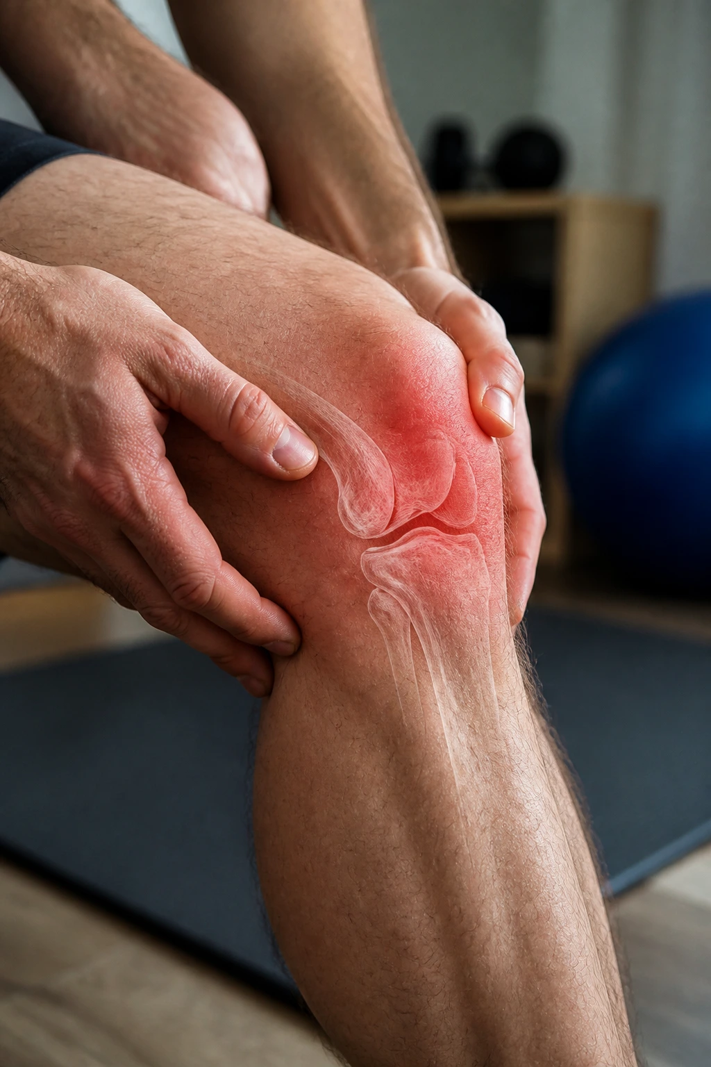
You have two C-shaped pieces of cartilage in each knee, acting as shock absorbers between your thigh and shin bones. A tear often happens when you twist your knee while your foot is planted firmly on the ground. Unlike ligaments, which have a decent blood supply, the meniscus is mostly avascular, meaning it struggles to heal on its own.
Symptoms to Watch For
- A sensation of the knee “catching” or locking up when you try to straighten it.
- Sharp, stabbing pain directly at the joint line (the space between the bones).
- Swelling that comes and goes, especially after activity.
Management Strategies
If the tear is minor, conservative management works. You prioritize reducing swelling first—ice, compression, and elevation. Then, you focus on strengthening the surrounding musculature. You cannot fix the cartilage itself, but you can build the muscle around it to take the pressure off the joint. Focus on high-repetition, low-weight exercises like terminal knee extensions and seated leg extensions to keep the joint moving without grinding. If the knee is constantly locking, however, you need to consult an orthopedic specialist to see if a surgical “trim” or repair is necessary.
4. Medial Collateral Ligament (MCL) Sprain
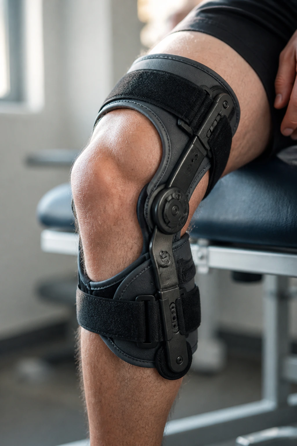
The MCL runs along the inside of your knee. It is often injured by a direct blow to the outside of the knee, which forces the joint to bend inward, stretching the inner ligament. Unlike the ACL, the MCL has a fantastic blood supply, which means it is often able to heal without surgery, even if it is a complete tear.
Assessment and Care
Most MCL sprains are graded on a scale of one to three. A Grade 1 is a minor stretch; a Grade 3 is a complete rupture. The good news is that for most people, rest and a hinged knee brace are enough to let the fibers knit back together.
- Use a Hinged Brace: This prevents side-to-side (valgus) motion while allowing the knee to bend back and forth.
- Avoid Pivoting: For the first few weeks, stick to straight-line walking. Lateral movements will re-stress the damaged tissue.
- Gradual Reloading: Start with stationary biking once the pain is manageable. The circular motion is gentle on the MCL compared to the shearing forces of running.
5. Patellofemoral Pain Syndrome (Runner’s Knee)
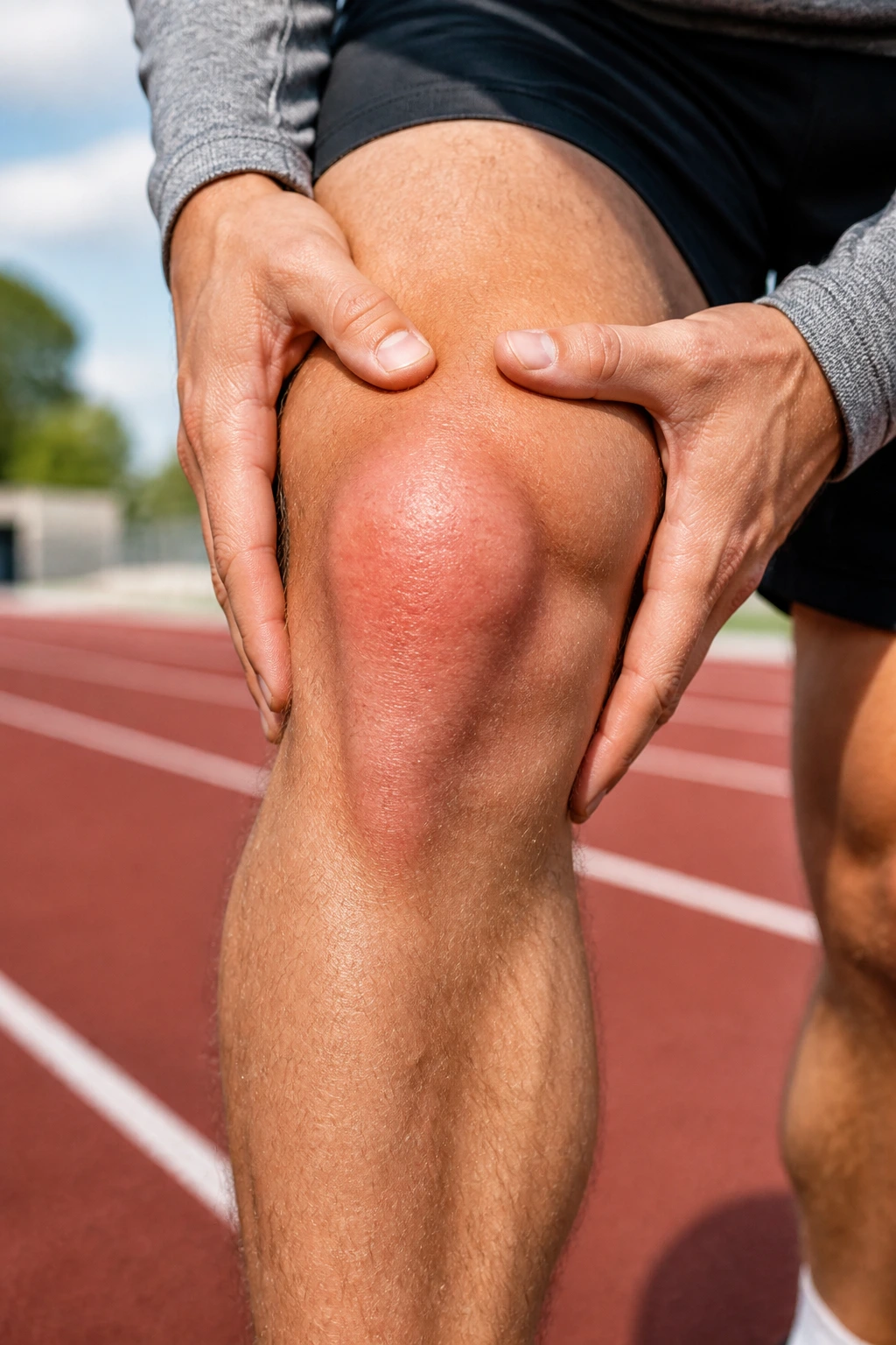
Runner’s knee is a broad term for pain around the front of the kneecap. It happens when the patella does not track correctly in the groove of the femur. Instead of gliding smoothly, it rubs against the bone, causing inflammation.
The Source of the Issue
It is almost always a tracking problem caused by muscle imbalances. If your outer thigh (vastus lateralis) is pulling harder than your inner thigh (vastus medialis), or if your hips are weak and causing your knees to cave inward, the kneecap won’t sit right.
Exercises for Correction
- VMO Activation: You need to specifically target the VMO (the teardrop-shaped muscle on the inside of the knee). Perform seated leg extensions, but focus on the last 15 degrees of extension—that’s when the VMO is most active.
- Hip Abduction: Weak glute medius muscles lead to “knee valgus” (knees caving in). Use resistance bands around your knees while performing side-steps or monster walks.
- Foam Rolling: Loosen up the IT band and the quads. Tight tissues around the knee pull the patella out of its proper groove.
6. Iliotibial (IT) Band Syndrome
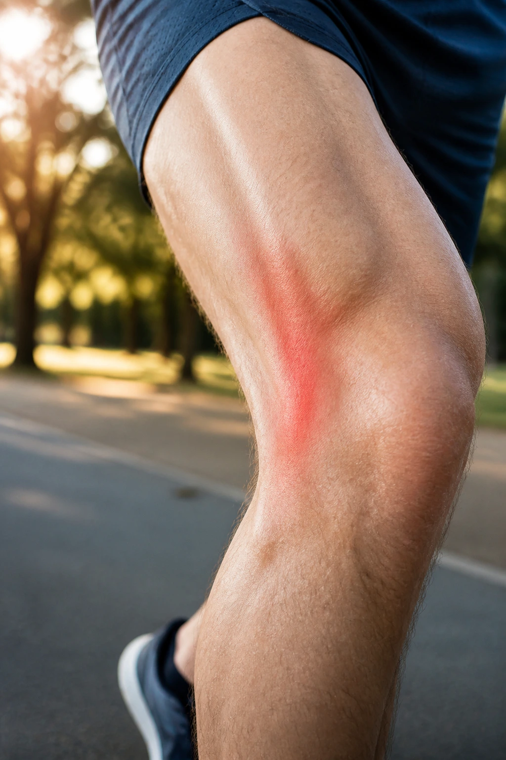
The IT band is a thick strip of fascia that runs from your hip down to the outside of your knee. When it becomes tight or inflamed, it rubs against the bony prominence on the outside of the knee, causing a sharp, stinging pain that usually kicks in after a specific distance during a run.
Why Stretching Doesn’t Always Help
Many people try to stretch the IT band by doing cross-leg stretches, but the IT band is dense, fibrous tissue—it doesn’t “stretch” like a muscle. You cannot loosen it just by pulling on it. Instead, you need to treat the muscles attached to it: the gluteus maximus and the tensor fasciae latae (TFL) at the hip.
Fixes that Work
- Glute Strengthening: Build the glutes. When the glutes are strong, they don’t rely on the TFL/IT band to stabilize the hip during a stride.
- Stride Length Reduction: Overstriding causes the heel to strike the ground far in front of the body, which puts massive stress on the IT band. Shorten your steps to keep your feet landing underneath your center of mass.
- Avoid the “Runner’s Pouch”: If you have hip inflammation, address that first, as it contributes to the tension running down the leg.
7. Knee Bursitis
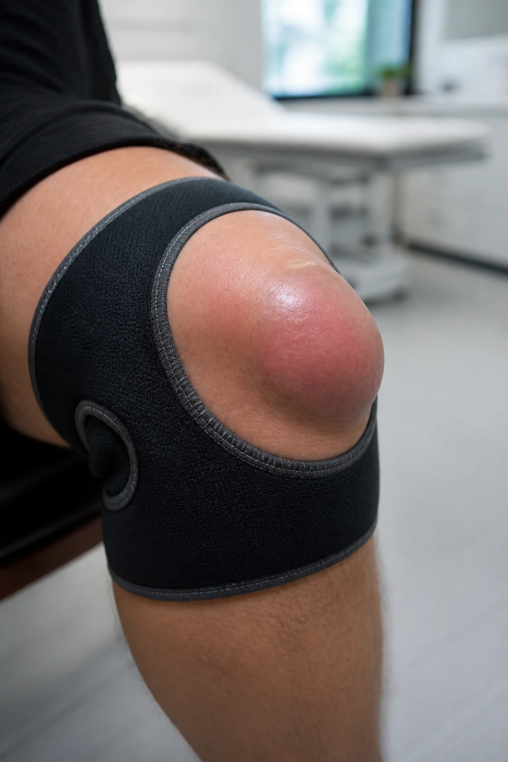
Bursae are tiny, fluid-filled sacs that act as cushions between your bones, tendons, and muscles. When these sacs become inflamed—often due to repetitive kneeling, direct impact, or overuse—you get bursitis. It feels like a localized, squishy swelling, usually right over the kneecap or slightly below it.
Management Techniques
- Cushioning: If you are in a profession that requires kneeling, use high-quality knee pads. Do not rely on your jeans.
- Compression: A compression sleeve can help reduce the swelling.
- Active Rest: Avoid the specific activity that caused the irritation. If it was lunging, switch to swimming or rowing until the bursa calms down.
- Anti-Inflammatory Protocol: Ice, elevation, and over-the-counter anti-inflammatories are usually very effective for bursitis, unlike structural ligament tears.
8. Chondromalacia Patellae
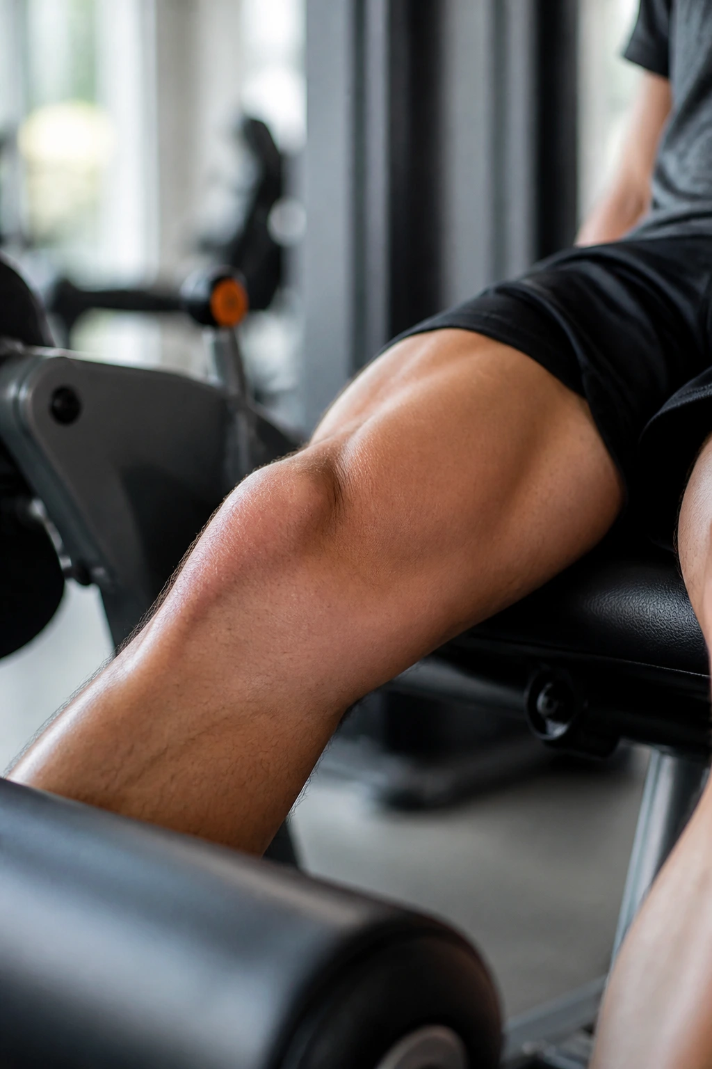
This is the softening and breakdown of the cartilage under your kneecap. It is most common in young, active individuals and is often referred to as “softening of the cartilage.” The primary symptom is a grinding or crunching sensation when you flex your knee, sometimes accompanied by a dull ache after sitting for long periods (often called “theater sign” because it hurts after sitting in a movie theater).
Addressing the Cartilage
Once cartilage is gone, it does not regrow. However, you can manage the pain and prevent further degradation by focusing on load distribution.
- Avoid Aggressive Deep Squats: If your kneecap is already grinding, doing heavy barbell squats will only accelerate the wear.
- Focus on Open-Chain Exercises: These are exercises where your foot is not fixed to the ground, like leg extensions. They can be performed with lower loads to build quad strength without the heavy compressive forces of a squat.
- Weight Management: Even small amounts of weight loss significantly reduce the pressure on the patellofemoral joint during walking.
9. Lateral Collateral Ligament (LCL) Sprain
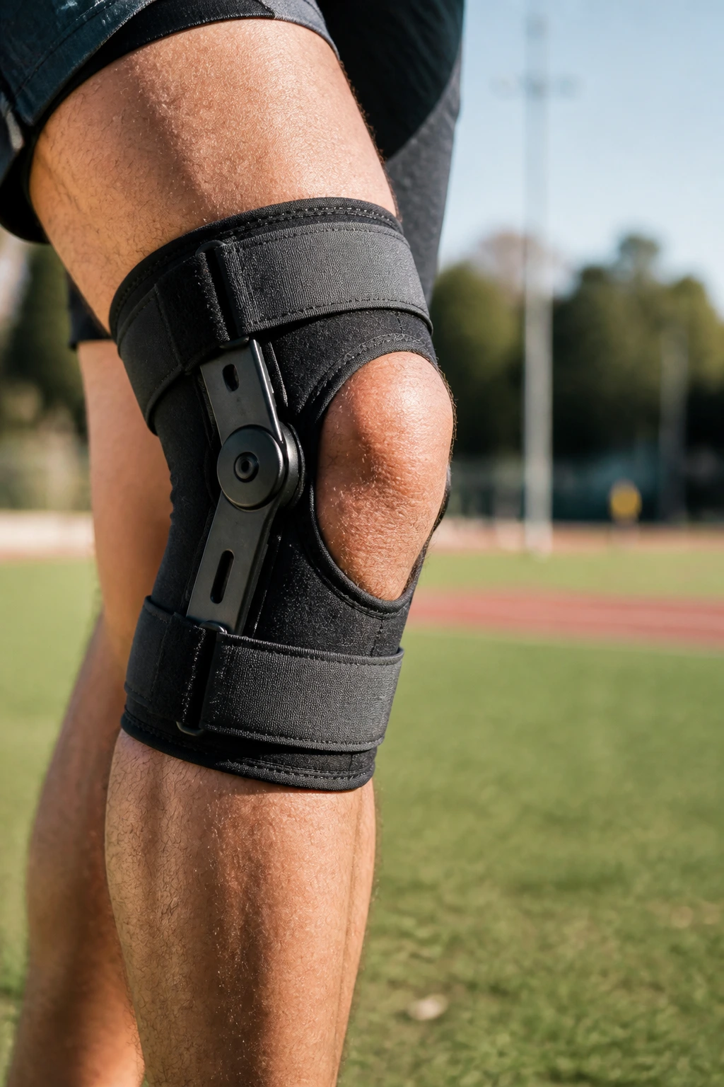
The LCL runs along the outside of the knee. Injuries here are much less common than MCL sprains because the structure of the leg naturally protects the outside of the knee from most forces. When it does happen, it’s usually from a blow to the inside of the knee that pushes the joint outward.
The Recovery Process
Because the LCL is often involved with other structures—like the popliteus muscle or the lateral meniscus—an LCL injury often requires a more thorough assessment than a simple sprain.
- Protect the Lateral Side: Use a brace to prevent varus stress (pushing the knee outward).
- Avoid Cutting/Pivoting: Just like with an MCL injury, you must avoid lateral movements until the ligament is no longer tender to the touch.
- Strengthen the Lateral Chain: Once pain-free, ensure your lateral stabilizers (hip abductors) are strong to prevent future outward “buckling” or instability.
10. Posterior Cruciate Ligament (PCL) Injury
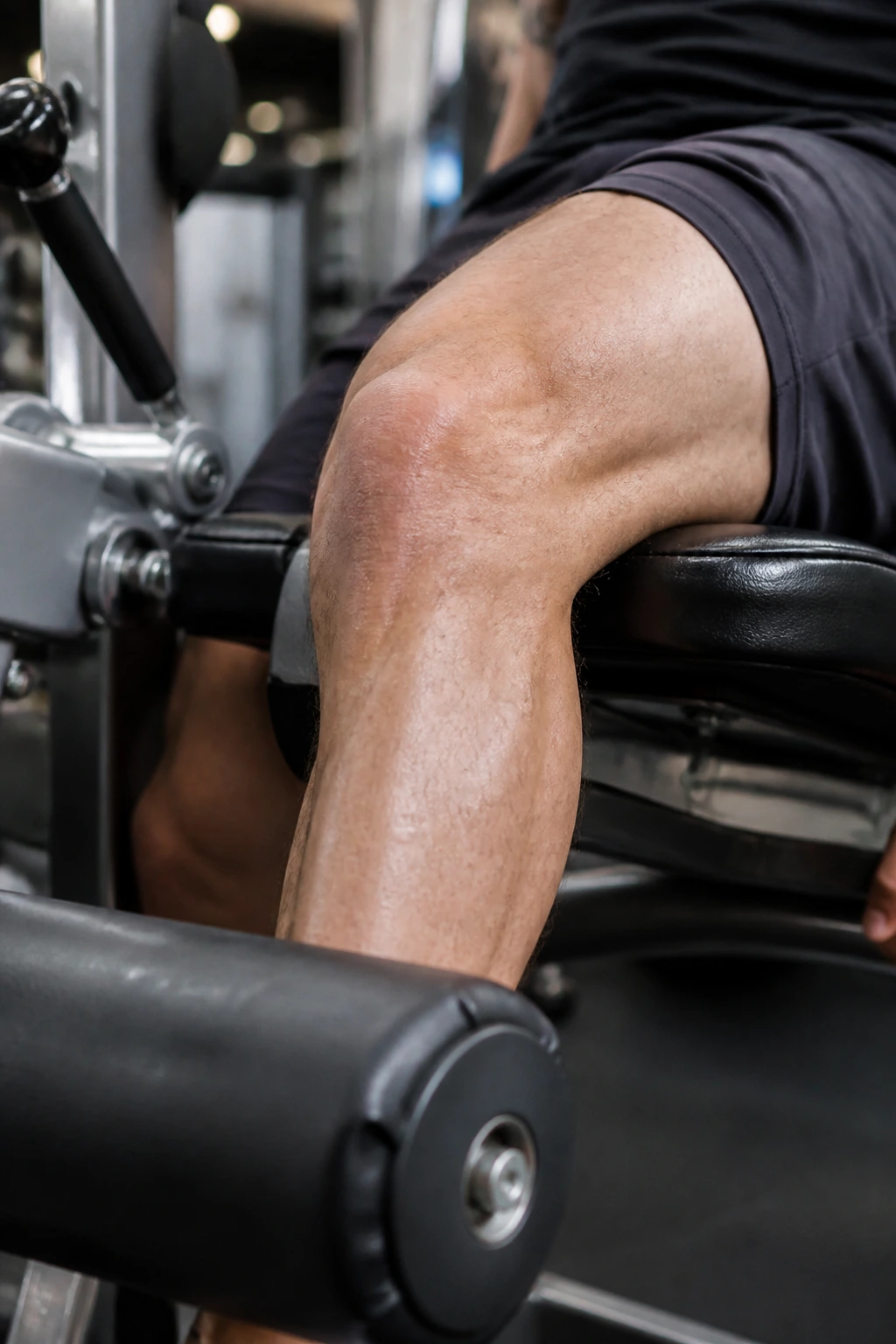
The PCL is the strongest ligament in the knee. It prevents the shinbone from moving backward under the femur. It is rarely injured in normal athletics; it usually takes a significant, direct blow to the front of the shin while the knee is bent—like hitting your dashboard during a car accident or falling hard on a bent knee in contact sports.
Healing and Prognosis
PCL injuries are rarely surgical. Because the ligament is so robust, it often heals well with conservative management.
- Quadriceps Focus: Since the PCL stops the shin from sagging backward, you want to build the quads to pull the shin forward, keeping the joint centered.
- Avoid Hamstring Dominance: The hamstrings pull the shin backward, putting more stress on the PCL. During the first phase of rehab, prioritize quad-dominant exercises over heavy hamstring loading.
- Patient Loading: PCL healing takes time. Do not rush back to contact sports.
11. Osteoarthritis
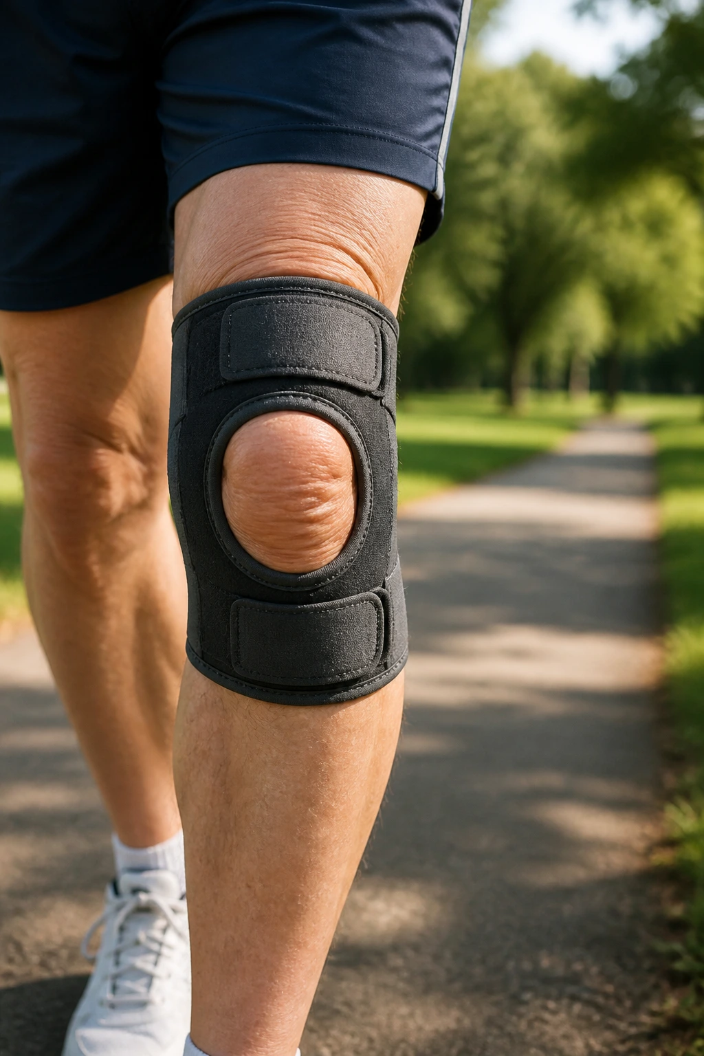
This is the wear-and-tear condition where the protective cartilage on the ends of your bones wears down over time. It is not necessarily an “injury” in the acute sense, but it is a chronic condition that many people deal with as they age. Stiffness, swelling, and pain that feels worse in the morning are classic signs.
Moving Through the Pain
The biggest mistake people with arthritis make is becoming sedentary. Motion is lotion for the joints. When you stop moving, the synovial fluid stops circulating, the muscles around the joint atrophy, and the stiffness becomes permanent.
- Low-Impact Movement: Cycling, swimming, and walking are your best friends.
- Strength Training: Building muscle around the knee helps “unload” the joint. Strong quads and hamstrings act as a protective barrier, absorbing forces that would otherwise go straight into the arthritic bone.
- Supplements and Nutrition: While not a “fix,” maintaining healthy inflammation levels through a diet rich in omega-3 fatty acids and antioxidants can help manage systemic joint pain.
12. Plica Syndrome
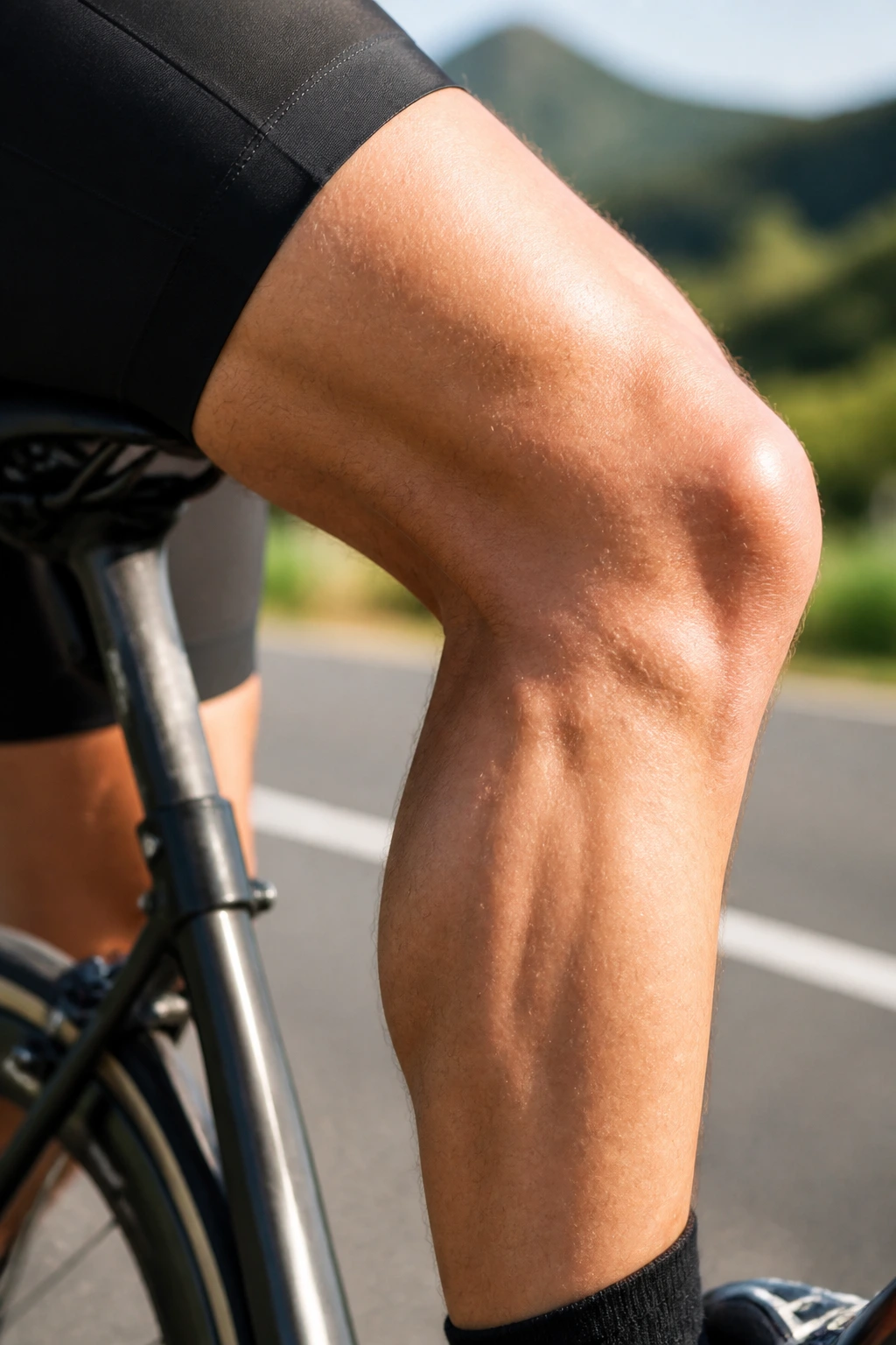
Your knee has a fold in the thin tissue lining the joint called a plica. If this fold becomes irritated or thickened, it can get caught between the bones, causing snapping, clicking, or sharp pain. It is often mistaken for meniscus issues because it feels like something is getting “caught” in the joint.
Managing the Snap
Plica syndrome is often worsened by activities that involve repetitive flexion and extension, like cycling with the seat too low.
- Adjust Your Equipment: If you are a cyclist, raise your seat. If you are a runner, check your cadence.
- Stretching the Quadriceps: Tight quads increase the pressure of the plica against the femur. Regular, gentle foam rolling of the quads can create more room for the joint to slide.
- Time and Rest: In most cases, if you avoid the aggravating motion, the plica will stop being inflamed and the symptoms will resolve.
13. Baker’s Cyst
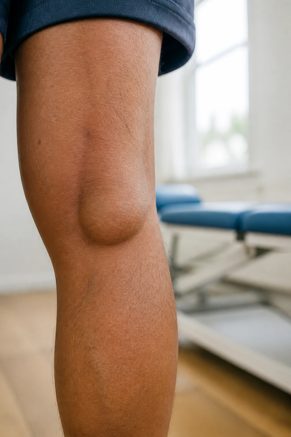
A Baker’s cyst is a fluid-filled swelling that causes a bulge behind your knee. It is rarely the primary problem; it is usually a symptom of something else, like a meniscus tear or arthritis, which causes the knee to produce excess synovial fluid. That fluid gets pushed into the back of the knee, creating the cyst.
The Real Fix
You can drain the cyst, but it will come back if you don’t fix the underlying cause.
- Identify the Cause: See a specialist to determine if you have an underlying cartilage or ligament issue.
- Compression: A knee sleeve can help manage the swelling behind the knee.
- Gentle Movement: Do not aggressively knead or massage the cyst, as this can rupture it, causing pain and swelling to spread down the calf.
14. Fat Pad Impingement
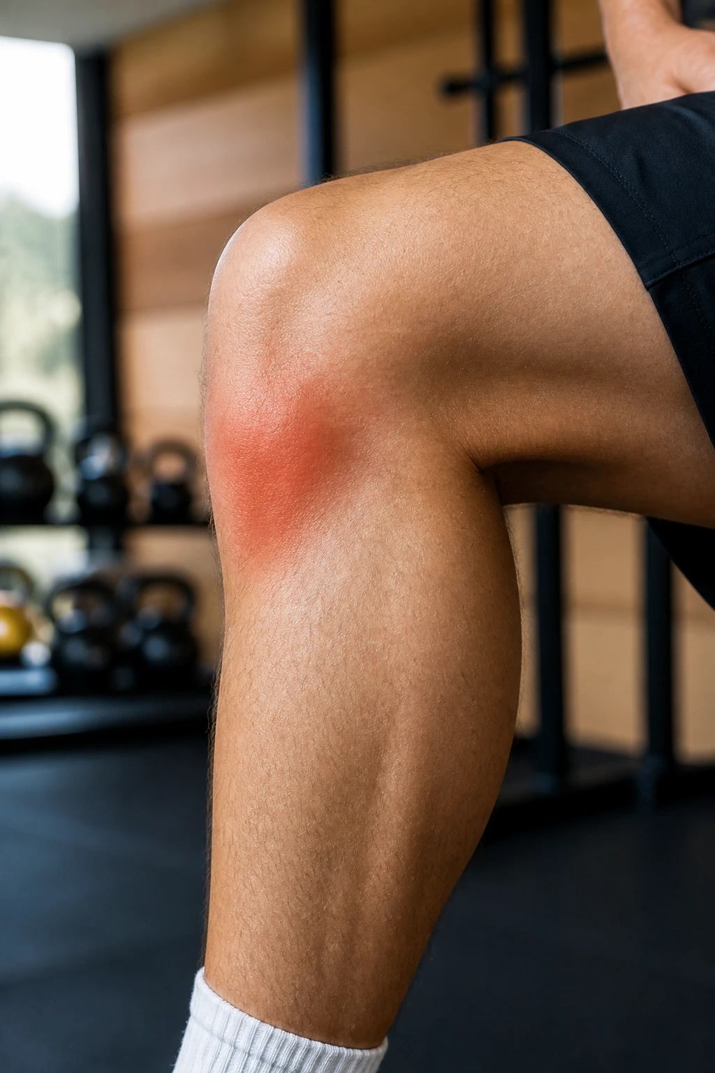
Right under your kneecap sits the infrapatellar fat pad, a highly sensitive area filled with nerve endings. If your knee extends too far or your kneecap is pushed out of place, this fat pad can get pinched, causing sharp, intense pain at the bottom of the kneecap.
The “Hyperextension” Problem
Fat pad impingement is very common in people who tend to stand with their knees locked in hyperextension.
- Train the Stop: Stop locking your knees. Practice standing with a “soft” knee, even if it feels unnatural at first.
- Taping: Sometimes, taping the patella slightly upward can help prevent the fat pad from getting caught.
- Avoid Over-Extension: In the gym, do not “snap” your knees at the top of a leg press or leg extension. Stop just short of full lockout.
15. Osteochondritis Dissecans
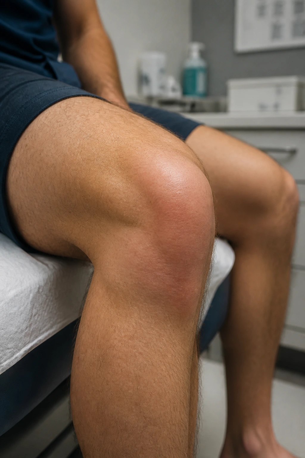
This is a condition where a piece of bone or cartilage loses its blood supply and dies. This can cause the piece of bone/cartilage to loosen and potentially float inside the knee joint. It is most common in adolescents, but it can occur in adults as well.
The Warning Signs
- Persistent, localized aching.
- Swelling that comes and goes.
- The sensation of something “floating” or locking in the joint.
Taking Action
This is not a “wait and see” injury. If you suspect your bone health is compromised, see an orthopedic surgeon immediately. Imaging—like an MRI—is required to see if the piece is stable or if it has detached. If it is stable, rest and immobilization can allow the bone to re-vascularize and heal. If it is loose, surgical intervention is usually the only way to remove or fix the loose fragment.
Final Thoughts
Healing a knee injury is an exercise in patience. It requires a fundamental shift in how you view your body—moving away from “pushing through the pain” and toward “listening to the mechanics.” Your knees are not disposable parts. They are high-functioning, intricate biological systems that respond to stress with either adaptation or breakdown.
The goal of recovery is to return to your baseline, but the “pro” move is to return stronger than you were before the injury occurred. By addressing the weaknesses in your hips, ankles, and core that likely contributed to the initial issue, you turn a period of rehab into a long-term strategy for resilience. Listen to your body, respect the timeline of your tissues, and trust the process of gradual loading. You will be back on your feet—and on the trail, court, or gym floor—before you know it.